Artificial intelligence (AI) has come to play an increasingly integral role in medical imaging in general, and in the field of urology particularly. The applications of AI in urology are numerous, starting with accurate diagnosis (using image segmentation and abnormality detection), continuing with biopsy and operative treatment (using tools for assisted navigation and robotic guidance), and ending in treatment assessment (using tools similar to those used in diagnosis in order to assess the response to treatment).
Imaging in urology
The field of urology encompasses a wide variety of diseases that inflict the urinary-tract system, including the kidneys, ureters, bladder, and urethra. It also encompasses diseases that solely inflict the male reproductive organs in general, and the prostate gland in particular. People of all ages are inflicted with urological diseases, ranging from life threatening cancers to noncancerous health problems, such as benign prostate hyperplasia or kidney stones.
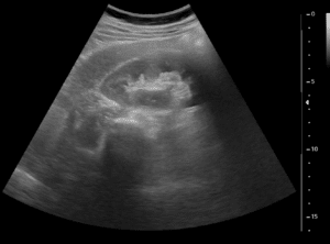
In recent years, medical imaging has come to play a central and essential role in both the diagnosis and the treatment of diseases in the field of urology. A wide variety of imaging techniques are used in order to accurately diagnose and treat patients suffering from urological diseases, including but not limited to microscopy, US, X-ray, CT and MRI.
The role of AI in urology
The increase in multipurpose and multimodal imaging techniques has amplified the need for quick and accurate image analysis and artificial intelligence tools. These tools are developed and used in order to effectively assist urologists in decision making, while increasing accuracy and reducing the time and cost spent in the process.
AI plays an important role in diagnosis, treatment, and assessment of response to said treatment in different urological diseases:
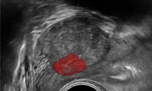
- Accurate diagnosis– tools such as image segmentation and abnormality detection are used for locating and grading tumors.
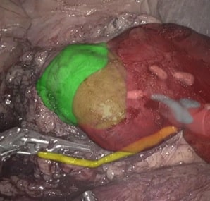
- Biopsy and treatment– during interventional procedures, tools such as 3D reconstruction, motion estimation, and action recognition are used for assisted navigation and robotic guidance.
- Treatment assessment– tools similar to those used during diagnosis are used in order to assess the patient’s response to treatment.
Current challenges
Image processing and AI pose many challenges in urology. Following are two significant challenges relating to treatment of prostate cancer that have been tackled by RSIP Vision:
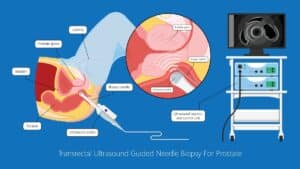
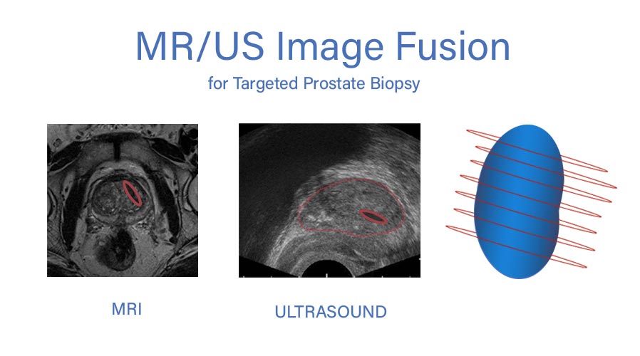
- Image fusion– In some cases, such as MRI-targeted-ultrasound-guided biopsies of prostate tumors, multimodal imaging techniques are used simultaneously. While MRI delivers high resolution images, its scanner time is limited, expensive and requires special tools. As a result, these prostate biopsies use previously taken MRI images during a live feed from US simultaneously.
RSIP Vision offers an alternative to manual registration (cognitive fusion): accurate, safe and robust software for quick and fully automated image fusion (registration). This assistance in the critical navigation during interventional procedures helps physicians ensure better overall clinical outcome.
- Video analysis– During some interventional procedures, small sensors are inserted in order to get a live video feed. For example, when a prostate gland is enlarged due to a malignant or a benign tumor, tissue often needs to be excised. The main challenge involves accurately reaching the targeted tissue using live video feed.
RSIP Vision has made significant progress in tackling this challenge by using video feed taken by a sensor in order to reconstruct a three-dimensional image. RSIP uses novel algorithms that combine classic computer vision and deep learning to complete the process. The result is better guidance and improved accuracy during interventional excision of prostate tissue.
While the challenges and solutions discussed here relate to urological diseases of the prostate, they most certainly also apply for other urological diseases, particularly those that demand interventional procedures. Biopsies of different kinds and treatment of kidney and bladder cancers demand better image analysis and AI tools for improved patient treatment and outcome.
A previous version of this article was published on Computer Vision News

 Urology