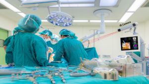
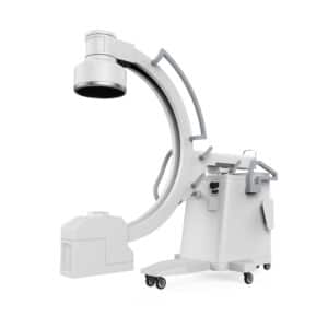
Fluoroscopy is an extremely useful imaging tool in surgical procedures in orthopedics, cardiology, GI, etc. In some cases, it is used as a real-time modality and it is a well-established method for image interpretation. Recent advancements in AI and computer vision technology also allow 3D reconstruction from these images, as well as fusion to other modalities, making it a better tool for navigation. The main detriment of fluoroscopy is the ionizing x-ray radiation which penetrate the human body and can cause long-term damage. Increased risk of cataracts as well as a variety of cancers, such as breast cancer, thyroid cancer, burns, are prevalent in all those involved in fluoroscopy guided procedures – surgeons, OR staff, and in some cases also the patients.
How to Lower Radiation Exposure?
Minimizing exposure to radiation for all involved in the procedure is essential. The precautionary principle used for this is ALARA – As Low As Reasonably Achievable – essentially aiming to use as little radiation as possible without compromising the procedure. One method to lower radiation exposure is low-dose imaging. This results in grainy images with low signal-to-noise ratio, making them difficult to decipher and lowering treatment quality.
Using Deep Learning to Improve Low Quality Images
There are two methods to use low dose images without compromising the quality of the images. The first one is to improve a single image using deep leaning state of the art methods.
The low-dose images have a characteristic grainy background and blurriness. State-of-the-art deep learning methods use convolutional neural networks to learn the transformation from the standard images to the low-quality images and vice versa. This is done by training the system with large data sets containing both types of images. Using this transformation can create a high-quality image from the low-dose image, improving the image quality significantly. This results in comparable image quality with a radiation reduction of up to 75%.
The second method is using the data that we have from other images. In many cases the fluoroscopy images are taken in series – this way we can use data from previous images to improve current images. Here deep learning can be used to sharpen and remove the noise artifacts from the images.
Lower Radiation in Your Device
RSIP Vision has extensive experience in implementing image enhancement algorithms in medical devices. Our multidisciplinary team of engineers and clinicians can quickly and efficiently execute a module which will improve image quality despite low dosage imaging, speeding time to market for your device. Contact us today!


 Surgical