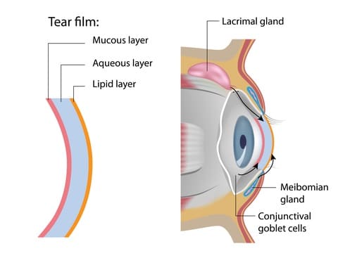
Infrared meibography is an imaging technique producing excellent quality images of meibomian glands, but not sufficient to provide information at the microscopic level. For this purpose, other technologies have been proposed, like the laser confocal microscopy, which uses laser scans to acquire very clear and precise images of the microscopic structures of the meibomian glands. However, this type of meibography is slightly more invasive, since it requires eyelid eversion: to decrease pain and facilitate procedure, topical anesthesia is applied before performing alignment of the lens against the posterior aspect of the everted eyelid.
Evaluation of Meibomian Gland Dysfunction
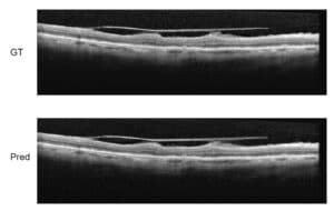
The advanced way to evaluate meibomian gland dysfunction is by using highly accurate OCT, which delivers a 3D tomography of the glands. It may display features and measurements which are crucial to understand the specific problem of each gland.
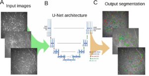
The image analysis of the gland is very complex, because what is seen is not one organ with one contour: it looks like a grape-like structure, which in the normal eye is composed of many small clustered parts. In the abnormal eye, the observer will see differences in the topography of the gland which need to be explained. Filtering techniques for noise reduction and thresholding allow to perform a correct segmentation of the gland. Fourier Transform is the main algorithmic technique enabling us to decompose the image and perform the image processing operations used to detect and describe the presence of meibomian gland dysfunction: this and other tools help to accurately assess the regularity of the gland and of its structure. RSIP Vision has decades-long experience in these advanced imaging procedures. Share with us your work and your projects!

 Ophthalmology