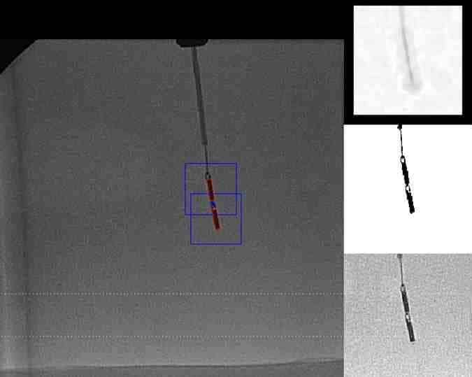
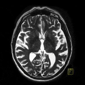
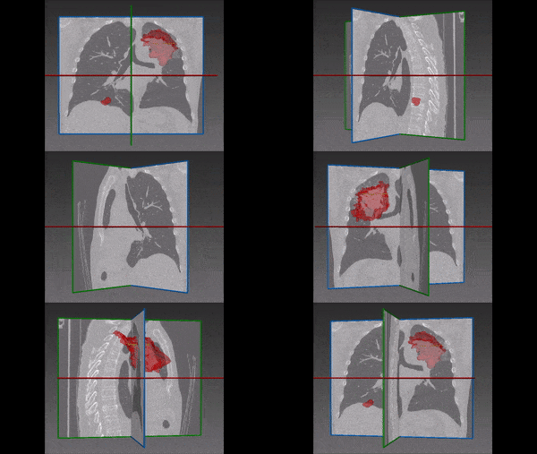
The localized MRI works by inserting rectangular shaped catheter (see image above) through patients’ vascular system and careful navigation up to the target vessel. At the target site, the catheter is carefully rotated on its axis to capture 360 image of the vessel. To construct the MRI image, the orientation of the catheter needs to be correlated to the image obtained at each moment of acquisition, to be able to construct the 3D vessel. However, fine trembling, patient motion and operator faults will result in the catheter not rotating smoothly and at equal speed, and therefore the sequence of acquisitions cannot be reconstructed to give a reliable 3D surface or panoramic image.
Magnetic Resonance Imaging software by RSIP Vision
For this end, RSIP Vision has constructed an image processing and computer vision-based algorithm, which can detect the orientation, speed and direction of motion of the rectangular catheter’s heads, as if continuously being moved by the operator. Challenges of low signal to noise ratio were successfully dealt with throughout the project, to accurately extract the orientation of the catheter. We then obtained a one-to-one correspondence between the orientation of the catheters and the acquired image and stitched the images in such a way that they match the correct spatial position of the catheter within the vessel. We thus generated a precise image of the fibrous or fatty tissues in a vessel.
RSIP Vision has been constructing tailor-made algorithmic solutions to the medical industry for over 2 decades. We put emphasis on meeting the accuracy constrains imposed by our clients, allowing them to surpass industrial and regulatory gold standard and push their product to the forefront of technology. To learn more about RSIP Vision’s projects in the medical field, please visit our project pages.

