Lymph nodes are routinely examined and assessed during physical examination of patients in a clinic or hospital setting. Enlarged lymph nodes can be indicators of infection, cancer and other pathologies. Therefore, a biopsy of a suspected lymph node, which provides tissue histology or cytology is a vital step towards diagnosis.
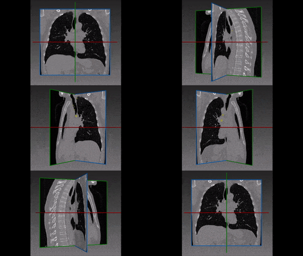
In relation to malignancy, initial staging before treatment is done using the Ann Arbor staging system or Lugano classification, by analyzing patient’s computed tomography (CT) and positron emission tomography (PET) scans. In addition, tracking the changes in lymph node’s size, is indicated during and after treatment e.g, radiation or chemotherapy. This monitoring may reflect treatment effectiveness. Again, the assessment of response can be done using the Lugano classification, and more recently by the lymphoma response to immunomodulatory therapy criteria (LYRIC).
Automated lymph node segmentation
Analysis of lymph nodes can be difficult and time-consuming for the radiologist. Hence, automatic detection and segmentation of lymph nodes from CT scans is important and valuable in order to eliminate errors. Automated lymph nodes segmentation requires expertise in differentiating lymph node boundaries from adjacent structures. An automatic algorithm would provide the physician with relevant information for and during the lymph node biopsy and also supply accurate information for diagnosis and management.
RSIP Vision built an advanced AI module, based on the cutting edge convolutional neural network architecture, for segmentation of lymph nodes in CT scans using deep learning algorithms. This module was fine tuned to withstand the various challenges encountered while trying to determine whether each voxel belongs to a lymph node or not and resulted in one of the quickest and most accurate ways to perform this task. If you are still performing this task manually, we have the ideal software solution for you. Talk to an RSIP Vision engineer now.

 Medical segmentation