However, also automatic left ventricle segmentation (LV segmentation) faces challenging difficulties, like locating the left ventricle and overcoming the lack of edge information. We recommend a method to provide fast, accurate and fully automatic location and segmentation of the left ventricle, which takes into account also papillary and trabecular muscles, as well as identifies endocardial and epicardial contours in all the cardiac phases.
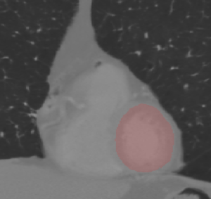
One of the main advantages of our recommended method is that it does not require any hand-made initialization, either by drawing manual contours or by adhering to a prior statistical heart shape model: the location of the left ventricle blood pool is automatically identified in the end diastole phase by the way of a roundness metric, under the assumptions that: the region of interest (the heart) is approximately in the middle of the image; that left and right ventricles’ blood pool have different circularity; and that blood has brighter signal intensity than the heart muscle. Another assumption is that the central part of the left ventricle blood pool does not move much across image slices and cardiac phases, which enables us to subsequently locate the contours of the blood pool, the papillary muscles (a small muscle within the ventricle that anchors the heart valves) and the trabeculations (pieces of muscle that extend into the ventricle). This is a crucial step, though it is sometimes neglected in other techniques of heart segmentation, since papillary muscles and trabeculations have a substantial impact on the measure of left ventricle volume, mass and ejection fraction1.
A region growing algorithm is used, after the image is converted to binary. Pixels are added in accordance with the intensity criterion (minimal difference in brightness). Image holes are then filled via morphological operations. Contours are finally smoothed with the help of a Fast Fourier Transform (FFT) technique, which removes all data points that are far from most other points of the dataset, without changing the overall shape.
The proposed method provides very robust input for daily clinical application, in line with expert-found values. Its only limitation to date is in the area of detecting contours in hypertrophic subjects. Future research will introduce additional constraints to take care also of this difficulty.
1 The health of the left ventricle can be (among other means) by quantitatively measuring its ejection fraction, stroke volume and ventricular mass, all critical parameters for cardiac diagnosis. Ejection fraction quantifies the percentage volume of blood spread out of the left ventricle in a given cardiac cycle. Stroke volume is the amount of blood pumped out of the left ventricle to the body.

 Cardiology