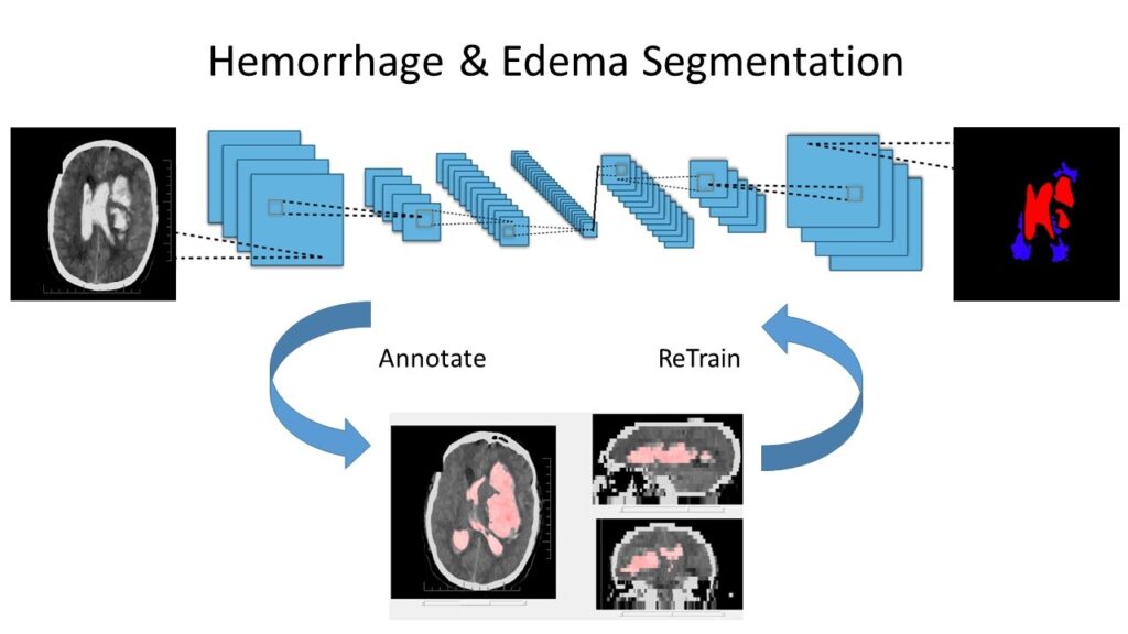
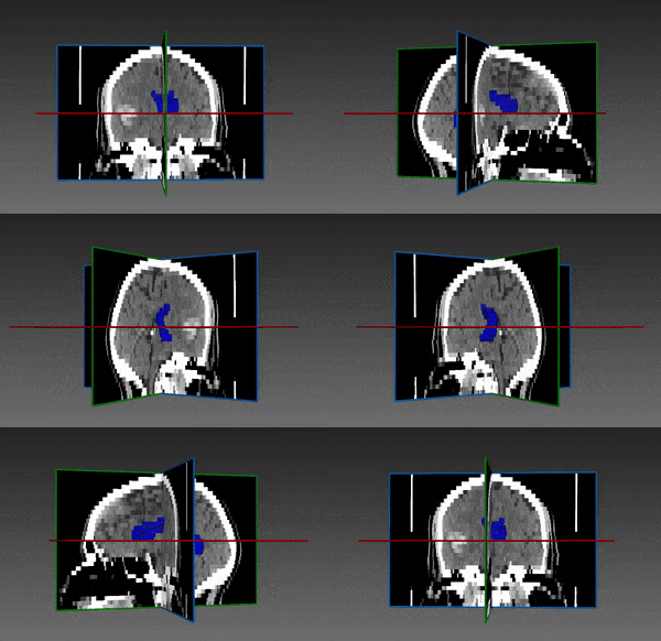
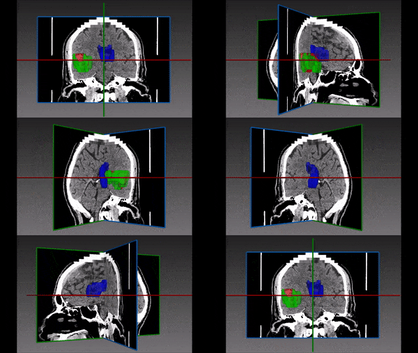
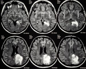
Edema is exceedingly difficult to identify, appearing as a subtle darker area surrounding the hemorrhage; it sometimes requires analysis of multiple sequential scans. Implementation and execution of a successful segmentation model in these situations requires expertise in all computer vision methods – both classical and deep learning based. RSIP Vision leverages both deep learning and classical computer vision techniques to provide a fully automated solution to this segmentation problem.
Fully Automated Intracranial Hemorrhage and Edema Segmentation
In order to do so, it is first necessary to solve the common challenge in medical imaging: the lack of labeled training examples, particularly for segmentation tasks. To solve it, our engineers started by using classical computer vision methods, such as graph cuts, superpixels and more, to quickly achieve a strong semi-automatic segmentation solution. This enabled an expert to quickly and easily label an initial training set, which was used to train a small neural network. Subsequently, we implemented a closed-loop bootstrapping solution to iteratively improve the training set, and train always bigger and better neural networks. As a result, RSIP Vision’s fully-automated segmentation technology achieves fast, robust and accurate segmentation of multiple cranial hemorrhage types, with constant running time.
Our hybrid artificial intelligence approach is effective and applicable when labeled training samples are limited or even nonexistent. Would you like to know how we can apply both classic computer vision and convolutional neural networks to your segmentation tasks? Contact us and our expert engineers will tell you how we can do it.

