The vision may be blurred due to the eye failing to achieve normal visual acuity. Strabismus becomes a problem which needs to be properly addressed when the newborn’s eyes fail to straighten and keep wandering by his or her 6th month of life. Pediatric strabismus is largely treatable with an appropriate and timely diagnosis. Early treatment improves the chances of developing good vision and proper depth perception.
Treatment for pediatric strabismus
Pediatric strabismus can be treated in several ways; sometimes, wearing eyeglasses is sufficient to straighten out the eyes. When it doesn’t, vision therapy uses an eye patch to cover the dominant eye forcing the weaker eye’s muscles and vision to fortify. Lastly, if the condition is not yet fixed, eye muscle surgery may be recommended. Therefore, early diagnosis is crucial.
Accurate diagnosis usually requires expensive equipment and an ophthalmology expert; the diagnosis involves cover tests or corneal light reflex tests. However, due to lack of experts and appropriate equipment, prompt diagnosis might not be easy to obtain. An excellent solution to the problem might be telemedicine with automated image analysis.
Image processing for strabismus diagnosis
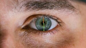
In order to diagnose strabismus from an image it is necessary to have both eyes in the image for relative comparison of orientation and alignment. The Hirschberg test checks the ocular alignment by corneal reflections: detection of strabismus is based on the location of first Purkinje, which are luminous reflections from the outer surface of the cornea. This information is obtained through an automated image processing task involving several steps: first, the precise locations of the eyes must be detected using algorithmic techniques that involve morphological features, position, threshold of grey level and shape. The second step is detecting the cornea, which is done by using some of the image processing methods also used in gaze tracking LINK: RANSAC (RANdom SAmple Consensus), fast radial symmetry transform and gradient filtering. These methods for detection of the iris circumference (the limbus) and its center presume a round object. The corneal light reflex is automatically detected by the grey levels around the center of the iris. Central corneal light reflex ratio (CCLRR), is then measured (taking into account the pupillary margin) and can be compared to normal and abnormal values to assess strabismus.
Another method would be to measure the distance between the corneal light reflex and the center of the pupil. The downside of this method is that detect pupils in dark eyes is far more difficult than in lighter eyes. RSIP Vision’s solid experience in image processing for detecting ophthalmology conditions recommends the use of the previously described solution. Our experts can write the best software for your eye care projects: contact them now.

 Ophthalmology