Skip to content
  • Our Work
    • Fields
      • Cardiology
      • ENT
      • Gastro
      • Orthopedics
      • Ophthalmology
      • Pulmonology
      • Surgical
      • Urology
      • Other
    • Modalities
      • Endoscopy
      • Medical Segmentation
      • Microscopy
      • Ultrasound
  • Success Stories
  • Insights
    • Magazine
    • Events and Webinars
    • News
    • Blog
  • The company
    • About us
    • Careers
Menu
  • Our Work
    • Fields
      • Cardiology
      • ENT
      • Gastro
      • Orthopedics
      • Ophthalmology
      • Pulmonology
      • Surgical
      • Urology
      • Other
    • Modalities
      • Endoscopy
      • Medical Segmentation
      • Microscopy
      • Ultrasound
  • Success Stories
  • Insights
    • Magazine
    • Events and Webinars
    • News
    • Blog
  • The company
    • About us
    • Careers
Contact

Hyperspectral Imaging for Robotic Assisted Surgery

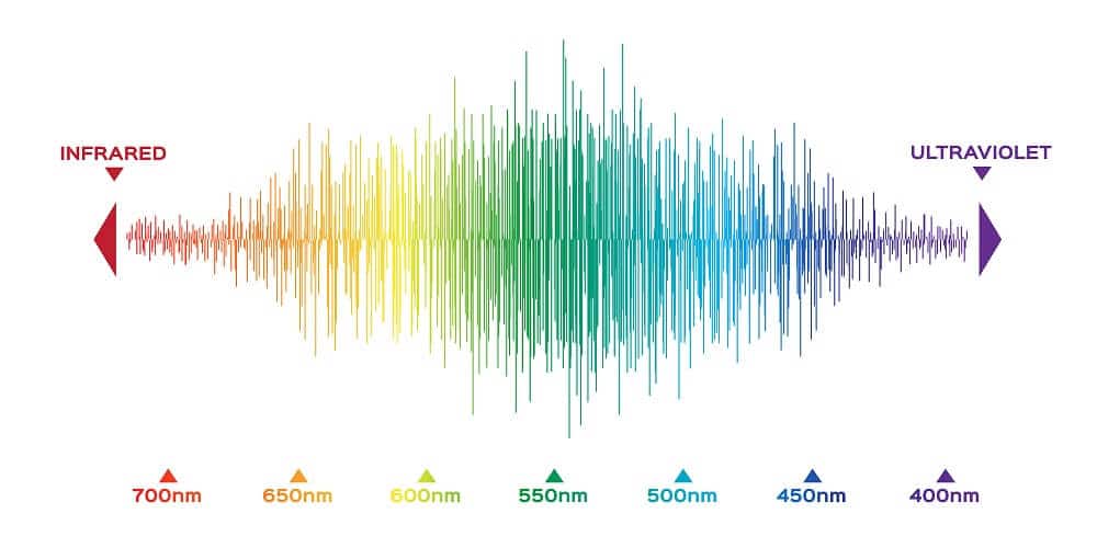
Standard imaging techniques make use of the visible light spectrum. As is known, the visible light is divided into three bands – red, green, and blue (RGB). This artificial division is very useful for visibility, however, pertinent information from different wavelengths is disregarded. Hyperspectral imaging utilizes this data to improve the visible image we see.

Visible spectrum color

How does it work?

Hyperspectral imaging requires that only a specific band from the spectrum will be recorded. This can be done by using a sensor that is sensitive to a unique part of the spectrum, or by adding a band pass filter before the sensor. It can be applied to several bands resulting in a large 3D array image in which the dimensions correspond to the x and y coordinate and given wavelength (A grayscale image is a 2D array where each pixel value is image intensity. An RGB image is equivalent to 3 grayscale images, or a 3D array where the wavelength dimension is 3). The value in each pixel in this array corresponds to the intensity of a specific wavelength. This image has significantly more data than standard images, making it harder to analyze. However, this data does not require neither contrast agents nor ionizing radiation.

Advantages of Hyperspectral Imaging

Materials have different reflection rates at different wavelengths. Standard imaging results in color differentiation of these materials, which is sufficient for visualization purposes. Hyperspectral imaging, on the other hand, has a better ability to differentiate between objects in the field-of-view, and even expose unseen objects. At specific wavelengths blood vessels beneath the surface will alter the reflection and therefore can be detected. Tumors have different material properties than healthy tissue, resulting in different hyperspectral reflection, allowing tumor recognition and segmentation. Hyperspectral imaging can detect differences which cannot be seen by the naked eye.

AI and Hyperspectral Imaging in Robotic Assisted Surgery

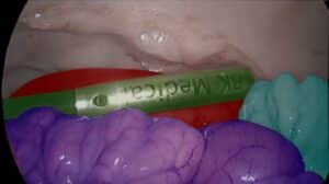
During Robotic Assisted Surgery (RAS), specifically in endoscopies, it is important to stay clear of underlying blood vessels and nerve bundles. Hyperspectral imaging can detect these in real time. Although the underlying structures can be detected, to have a full understanding of the anatomy below the surface, AI and deep learning methods are used. The underlying anatomy can be visualized by training the network to learn the hyperspectral data and segment the hidden blood vessels, nerve bundles, and tumors in the field of view. These can be portrayed on the RGB image and provide warnings to the surgeon, allowing to safely avoid or approach these structures, depending on the surgical needs. This capability adds another layer of safety to the procedure without using radiation (X-ray) or contrast agents.

Adding Hyperspectral Imaging to your Portfolio

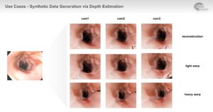
Analyzing hyperspectral data is challenging – it is difficult to find the relevant data in the “haystack” and doing so in real time requires very efficient algorithms. Our team of experienced clinicians and engineers utilize deep learning and other AI algorithms to train dedicated networks to automatically detect hidden objects of interest in real time using the hyperspectral imaging data and overlay them on the live image. This module has immense added value and can be immediately implemented into your medical device, enhancing a variety of use cases.

Share

Share on linkedin
Share on twitter
Share on facebook

Main Field

Surgical

In the recent years, robotic modules with their associated algorithmic software emerged in the surgery rooms. Here, some of their applications will be reviewed. They are used for orthopedic, brain and cardiac surgeries.

View Surgical

Categories

  • Robotics, RSIP Vision Learns, Surgical

Related Content

Single-port

How Can AI Assist in Single-port Robotic Surgery?

Data Generation in Robotic Assisted Surgeries (RAS)

Data Generation in Robotic Assisted Surgeries (RAS)

Success Rating and Dynamic Feedback in RAS

Success Rating in Robotic Assisted Surgeries

RAS Navigation

Tissue Sparing in Robotic Assisted Orthopedic Surgeries

C Arm X-Ray Machine Scanner

Radiation Reduction in Robotic Assisted Surgeries (RAS) Using AI

RAS Navigation

AI for Navigation in Robotic Assisted Surgeries

Show all

RSIP Vision

Field-tested software solutions and custom R&D, to power your next medical products with innovative AI and image analysis capabilities.

Read more about us

Get in touch

Please fill the following form and our experts will be happy to reply to you soon

Recent News

Announcement – XPlan.ai Confirms Premier Precision in Peer-Reviewed Clinical Study of its 2D-to-3D Knee Reconstruction Solution

IBD Scoring – Clario, GI Reviewers and RSIP Vision Team Up

RSIP Neph Announces a Revolutionary Intra-op Solution for Partial Nephrectomy Surgeries

Announcement – XPlan.ai by RSIP Vision Presents Successful Preliminary Results from Clinical Study of it’s XPlan 2D-to-3D Knee Bones Reconstruction

All news
Upcoming Events
Stay informed for our next events
Subscribe to Our Magazines

Subscribe now and receive the Computer Vision News Magazine every month to your mailbox

 
Subscribe for free
Follow us
Linkedin Twitter Facebook Youtube

contact@rsipvision.com

Terms of Use

Privacy Policy

© All rights reserved to RSIP Vision 2023

Created by Shmulik

  • Our Work
    • title-1
      • Ophthalmology
      • Uncategorized
      • Ophthalmology
      • Pulmonology
      • Cardiology
      • Orthopedics
    • Title-2
      • Orthopedics
  • Success Stories
  • Insights
  • The company