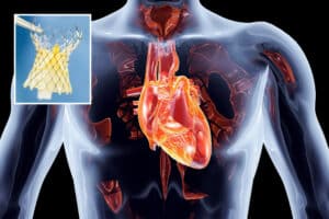
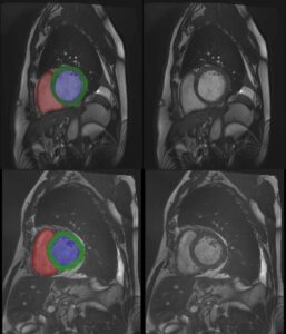
The great vessels conduct blood to and from the heart. These vessels include the aorta, superior and inferior vena cava, pulmonary arteries and pulmonary veins. Since the great vessels are an integral part of systemic and pulmonary circulation, vascular pathologies involving said vessels might be deadly.
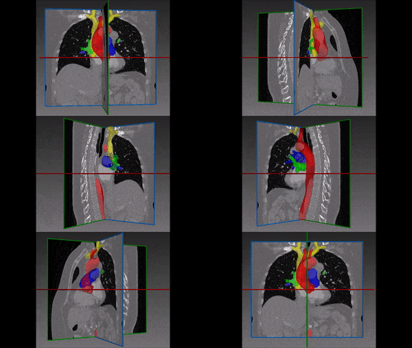
Computed tomography (CT) imaging is a useful tool for assessing the great vessels, and has become an essential tool for diagnosis of vascular pathologies, including aortic arch aneurysm, valvular heart disease and congenital vascular anomalies, such as aortic stenosis, coarctation of aorta or transposition of great vessels.
Furthermore, accurate imaging helps with surgical planning or endovascular procedures e.g. endovascular aneurysm repair (EVAR) or transcatheter aortic valve implantation (TAVI). Furthermore, it might even aid with quick diagnosis and surgical planning during life threatening vascular medical emergencies, such as aortic dissection or ruptured aortic aneurysm.
Moreover, by identifying vessels within the mediastinum, the physician can easily distinguish between vessels and other important structures including the heart, esophagus and trachea, or even abnormal masses. Hence, 3D segmentation of the great vessels proves to be extremely helpful as a diagnostic tool, surgical planning and guidance tool, as well as for follow-up.
Automated Great Vessels Segmentation:
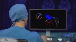
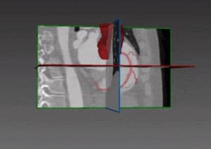
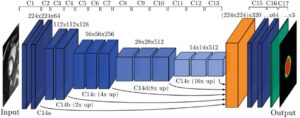
Utilizing deep convolutional networks, RSIP Vision’s engineers built a cutting-edge AI module for automatic segmentation of the great vessels. Combining global context and local information from the mediastinum area, the module is able to overcome the challenge posed by the target’s high anatomical variability. RSIP Vision’s fast and accurate module does not require any human intervention – making it the ideal instrument for identification and segmentation of great vessels in CT scans. Call us to discuss your cardio segmentation plans and take us along for your Deep Learning projects!

 Cardiology