General Medical

Annotation strategy and workflow

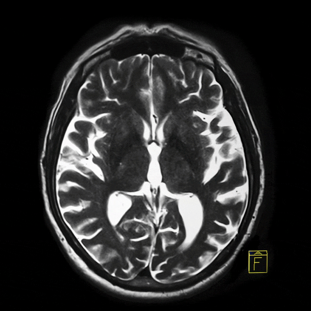
Data and Annotation Challenges in Medical AI Development

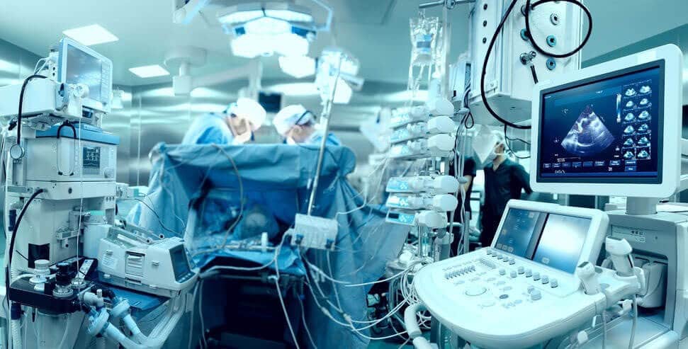
David Menashe, Arie Rond, Ilya Kovler Introduction In recent years, artificial intelligence (AI) has revolutionized many fields and has had a major impact on our daily lives. This revolution is driven by a combination of advanced deep learning models and an abundance of accessible training data on the internet. In the medical field, despite its …

Data and Annotation Challenges in Medical AI Development Read More »

Visualization Modalities for Surgery with AI

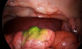
Visualization Modalities for Surgery with AI

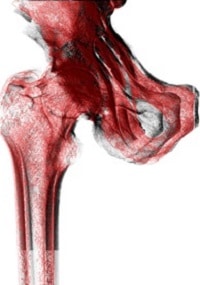
Our team has followed with deep attention the recent Hamlyn Symposium. In particular, we have enjoyed the great presentation by Professor Laura Marcu from UC Davis about fluorescence imaging during surgery, using that modality as a surgical aid. Fluorescence imaging is very widely used in microscopy: you inject a specific dye into the specimen, and …

Visualization Modalities for Surgery with AI Read More »

Navigation Systems for Surgery

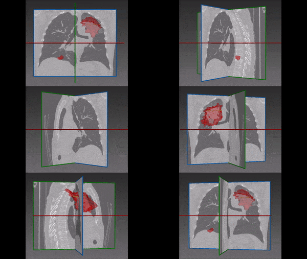
Catheter Navigation System with RL

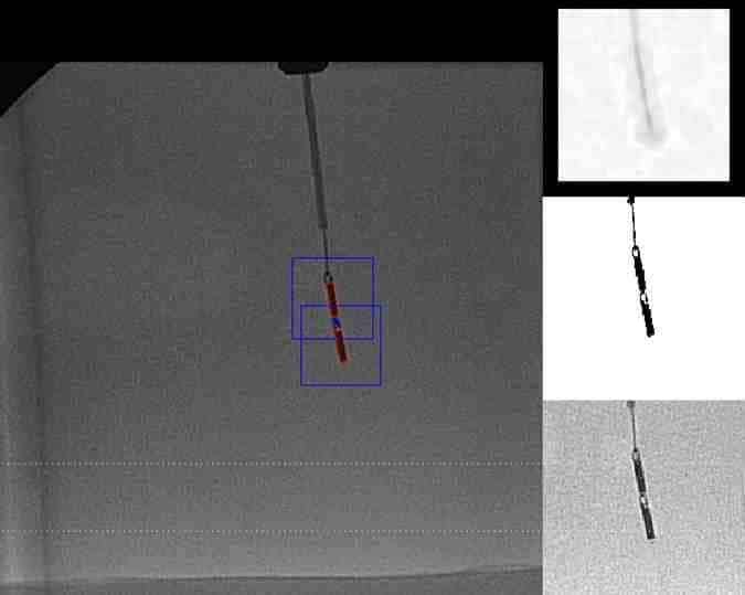
The typical catheter navigation system relies on fluoroscopy, which exposes patients to dangerous irradiation. To limit the dose throughout the operation, endoscopic cameras are used to guide catheter near the target site of the procedure. When vessel diameter becomes too small to insert an endoscopic camera, magnetic resonance imaging (MRI), CT or ultrasound scans are …

Catheter Navigation System with RL Read More »

U-Net network architecture

Deep Learning in Medical Imaging

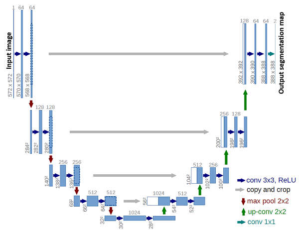
Until only a few years ago, traditional computer vision techniques have provided excellent results to detection and segmentation task. More recently, with the advent of deep learning  and neural networks also in medical imaging, we obtain surprisingly better results in all task, be it detection, segmentation, classification and the like. In this article we review the state-of-the-art in the newest model in medical image analysis.

Distorted Scan

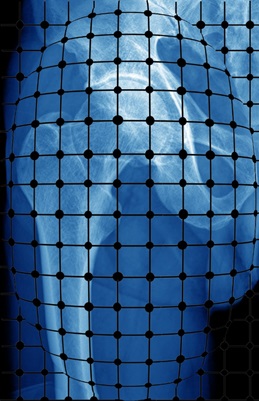
Calibration of X-ray images

This series of 5 articles by RSIP Vision about orthopedic navigation during surgery displays the state-of-the-art image processing techniques offering the surgeon a highly accurate and effective real time in-op view of the surgery environment. This procedure can be divided in several tasks: camera calibration of input imagessegmentation during orthopedic surgeriesregistration of CT and X-Ray; orientation and navigation during surgeryvalidation of accuracy in navigation systems. The outcome is a breakthrough advancement in orthopedic surgery performance, corroborated by rich academic literature supporting the method and by widespread use in the operation room.