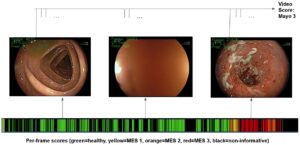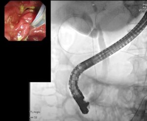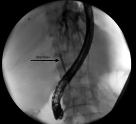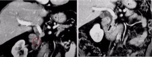
When imaging conditions permit relative clear field of view, such as in the case of endoscopic gastrointestinal examination, visual information can be used as a basis for computer vision algorithms working in real-time throughout diagnosis. Algorithms for the detection of lesion and other abnormalities can be tailor-made to meet accuracy demands and the expected anatomical position; they can also integrate patient-specific prognostic information for better detection. Trained eye and expert knowledge are critical requirements to distinguish a lesion from other anatomical structures. Such expertise is preferably translated into the algorithm’s code for automatic detection. However, difficulties pile up fast, due to incomplete ability to break down the steps required for us, as human, to distinguish one complex object from another.
Luckily, computer vision and machine learning put a large arsenal of techniques at our disposal. Clever utilization of these techniques enables to do just that, that is, translating expert knowledge into a fine-tuned algorithm, specifically designed for the task of lesion detection.
While probing the gastrointestinal, recognition of lesions relies on characterizing regions of abnormal color, reflectance, texture and shape. Segmentation of the plane-of-site into non-overlapping homogeneous regions, in terms of the aforementioned features, allows better characterization of the viewed image. In a non-predictable environment, obtaining satisfactory segmentation is non-trivial. It therefore requires an adaptive methods for segmentation: one such algorithm is the mean-shift segmentation algorithm.
Employing mean-shift to segment an input signal (image) requires sampled points in order to estimate their non-parametric density gradient. The mean-shift is an iterative algorithm, which shifts each sampled point to the weighted mean of the sample until convergence. Convergence is determined when the magnitude of the mean-shift vector reaches a predefined threshold value. In practice, image features form a cloud of points in some high dimensional space, for which the mean-shift algorithm is used for their clustering. The feature vector can include 3D color information, texture, neighboring information, local histogram, reflectance at each point, and the like.
Segmentation with the mean-shift requires to specify a kernel function (usually a radially symmetric), which governs the bandwidth (scale), i.e. the features weighting function. Features falling within the same scale are grouped to lay within the same spatial region of the image. Post processing steps are oftentimes required to merge over-segmented regions by removing erroneous borders to form more homogeneous regions.
Machine learning techniques are extremely useful at this stage to classify each region of the image. A sequence of controlled supervised classification is presented to a classifier (e.g. Support Vector Machine) until image region classification is reproducible to a satisfactory degree. Since a classifier is trained off-line, its performance for endoscopic examination can be made adequate for real time examination.
Taken together, adaptive segmentation of the image, using non-parametric methods, coupled with machine learning classification can be of a service in endoscopic examination. Such automatic tools can further be adapted to take into account relevant, patient-specific, medical information to help increase the accuracy of detection and decrease false ones, sparing further invasive procedures.
For computer algorithm to be integrated in medical devices and procedures, the highest level of robustness, accuracy, and reproducibility is required. Biased or lack of human expertise should not compromise patients safety during medical procedures, hence automatic methods can perform as a proper alert system for lesions. To comply with the stringent requirement which are necessary in medical devices, all project need a perfect expertise of these and other computer vision techniques. Be sure to always consult RSIP Vision’s engineers before you commence your project.

 Gastro


