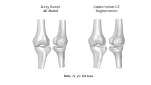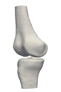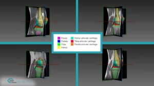
The extent of the intervention is determined by physicians based on the current erosion state. The state of erosion can be observed by the way of non-invasive imaging techniques such as simple ultrasonic imaging. Trained orthopedics evaluate the level of the intervention needed and steer the course of action based on, among other things, the thickness of the knee cartilage, as assessed by the physician. In fact, output images are analyzed and measured by hand or with semi-automated computational tools.
Automated density measurement of damaged cartilage
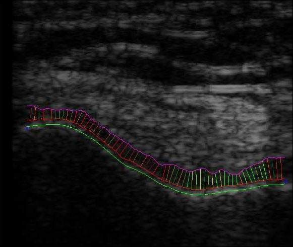
RSIP Vision has developed an automated tool to measure knee cartilage thickness based on ultrasonic images. Our automated algorithmic procedure releases physicians from the time-consuming human assessment. Ultrasound images of cartilage are hard to work with since borders between tissues are fuzzy. These boundaries take a cloud-like form, which can morph in both density and texture. To ensure automatic and accurate recognition of the different textures, we utilized active contour – ‘snakes’ technology to determine the borders of the cartilage. Our density measurement of damaged cartilage software is used by medical staff to produce a non-invasive, efficient and accurate measurement of knee cartilage density from ultrasound imaging of speckled cartilage tissue.
When shock absorption properties of cartilage are reduced by damage, RSIP Vision’s software enables orthopedics to assess state of erosion and decide treatment. To learn more about the activity of RSIP Vision in non-invasive orthopedic surgery, please visit our orthopedic surgery and medical segmentation project pages.

 Orthopedics