Cardiovascular disease at a glance
Cardiovascular disease is a leading cause of death, disability and health care expenditure in the United States.
- Cardiovascular disease (CVD) accounts for 17.9 million deaths worldwide each year, an estimated 31% of all deaths
- Nearly 30.3 million American adults are currently diagnosed with cardiovascular disease
- Cardiovascular disease and stroke cost more than $350 billion every year
Echocardiography
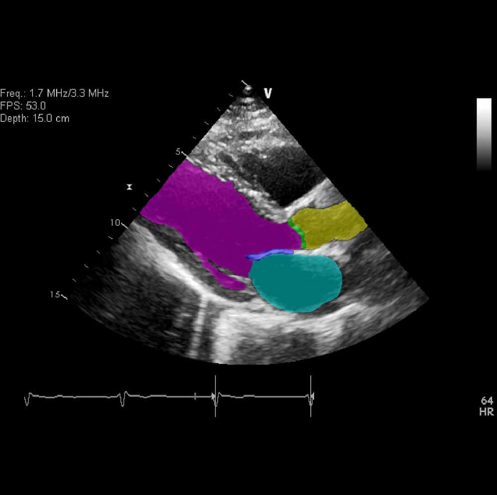
Cardiac ultrasound, or echocardiography, plays a crucial role in the diagnosis and management of cardiovascular disease. Echocardiography is the only imaging modality with real-time imaging of the heart, allowing immediate detection of pathologies and giving it a place across the entire cardiac patient care pathway. From pre-hospital care, the emergency room, general and cardiac intensive care, in the catheterization laboratory and operating room, echocardiography is fundamental.

Echocardiography uses ultrasound reflections of cardiac structures to generate images of the heart and vessels. These multiple images are then analyzed for anatomical anomalies and to evaluate possible pathologies. Several cardiac ultrasound technologies are available: Doppler analysis, M-mode echocardiography, two-dimensional trans thoracic echocardiography (TTE), trans esophageal echocardiography (TEE), and 3-D echocardiography. Cardiac ultrasound is safe, fast, non-invasive (apart from TEE) and does not expose patients to radiation. However, this staple of medical imaging is not without challenges.
Challenges in Echocardiography
Echocardiography is an important tool in the diagnosis and treatment of serious cardiac illness. Currently, ultrasound imaging processing faces two main challenges:
1. Image quality
Echocardiogram technicians must identify an ideal pathway for sound waves to pass in and out of the body. This ‘thoracic window’ must be relatively free from interfering obstacles and identifying this window requires precision and expertise. Height, body mass index, lung health, and body position must all be considered.
Lung disease and scar tissue from thoracic surgery can compromise images, and patients with limited mobility who cannot be rotated laterally also pose a challenge for obtaining an accurate echocardiogram. This difficulty was illustrated in one recent study that found echocardiographic quality inadequacy in 24% of imaging studies.
2. Image assessment
In light of operational challenges to image quality, image interpretation likewise poses a challenge. Studies have shown echocardiographic assessment inaccuracy may be as high as 30%. Traditional imaging interpretation relies on skilled physicians and researchers examining echocardiograms for pathologies. Interpreting images requires extensive training and practice, making it a complex and potentially costly and time-intensive process. Interpretation further relies on the subjective expertise of the human operators, which can lead to variability and error. This is especially problematic when interpreting lower quality images – a not uncommon outcome in echocardiography.
Given the centrality of accurate and rapid assessment of cardiac ultrasound images for effective treatment protocol and medical decision making, it is imperative to harness automation technology and advanced AI and image analysis technologies, in order to improve imaging quality and interpretation.
Addressing Challenges with Deep Learning for Echocardiography
Artificial intelligence is revolutionizing medical imaging across numerous modalities and is having a profound impact on cardiac ultrasound. Deep learning is a function of AI machine learning composed of hierarchical layers of algorithms mimicking the brain’s neural processes and designed to train itself to perform tasks. By learning features from examples, it then detects deviations from patterns in the data. The more training examples are included, the more efficient it becomes.
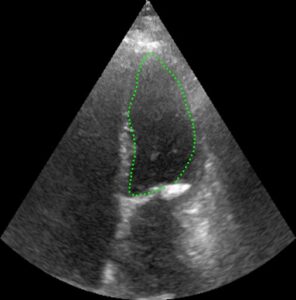
Deep learning is implemented in studies and in clinical use for detection, classification, segmentation, report generation and tracking in echocardiography. The direct application of neural networks will dramatically improve patient care, from primary identification to disease management, and address challenges to streamlined cardiac care. Advanced computational processing is fast and accurate, thereby lowering the cost of echocardiography while providing consistent, accurate and automated interpretation of echocardiograms. In one recent study, AI applied to over 14,000 echocardiograms demonstrated comprehensive efficacy in image classification, segmentation and diagnosis.
Deep learning is also used to enhance image quality, making it easier for physicians and researchers to interpret the images accurately. Additionally, deep learning for echocardiography is utilized to process and sort large amounts of imaging-generated data that would otherwise remain underutilized.
Another application involves training a deep learning model to explore the relationship between electronic medical records and cardiac imaging, thereby providing clinicians with insights to make more informed decisions in real time. By dramatically reducing the time required for cardiac diagnosticians to access and evaluate patient information in conjunction with echocardiography, AI can profoundly impact patient outcomes.
In a rapidly expanding market of artificial intelligence for medical applications, deep learning for echocardiography demonstrates significant potential in streamlining diagnostic workflow and reducing computational burden while improving quantification and accuracy in cardiac imaging. An additional and important effect of enhanced echocardiography is the democratization of advanced cardiac care. By simplifying and reducing the cost of cardiac imaging with artificial intelligence, patients in underserved areas can be treated with greater efficiency.
RSIP Vision: At the Heart of AI for Cardiac Care
RSIP Vision is resolving user variability, accuracy and efficiency in cardiac ultrasound with our advanced, deep learning based solutions. With applications spanning from prevention screening to real-time intervention guidance and assistance, RSIP Vision leverages advanced algorithms and image processing software to assist physicians in treating patients. Cardiac imaging is currently moving towards expanded integration of various AI solutions. This novel and efficient technology is transforming the utilization of deep learning for echocardiography in clinical pathways, making cardiovascular healthcare safer, more accessible and cost-effective. RSIP Vision is a leader in deep learning for echocardiography, at the heart of AI for cardiac care. Click here for more information about our computer vision solutions for cardiology.

 Ultrasound