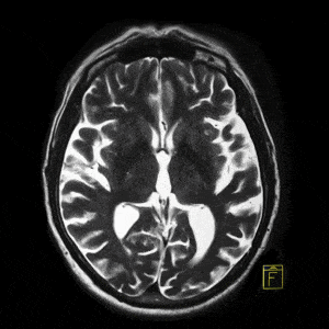
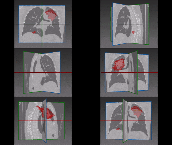
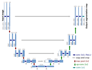
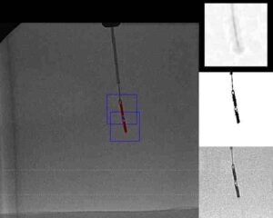
During the last few decades, the field of biomedical imaging was shaken by major breakthroughs, which have completely changed the way physicians can observe imaging data. For instance, not long time ago, radiologists received images (taken by CT, MRI or X-Ray) and had to analyze them from scratch. In the case of a full body MRI, they had to go through all the details of the body to check for any possible anomaly.
Needless to say, the process was very long and tedious; as such, it also generated mistakes. Modern computer vision technologies in biomedical image analysis enabled major progress in these procedures: for instance, the detection of lesions and grading of tumors or other pathologies are now performed with a level of accuracy close to that of biopsy, which is an invasive procedure. The result of this biomedical image processing is that radiologists can be alerted to what they are likely to find in the patient’s images. Of course, fastening the diagnosis can reduce the time to treatment, drastically improving its effectiveness on the patient.
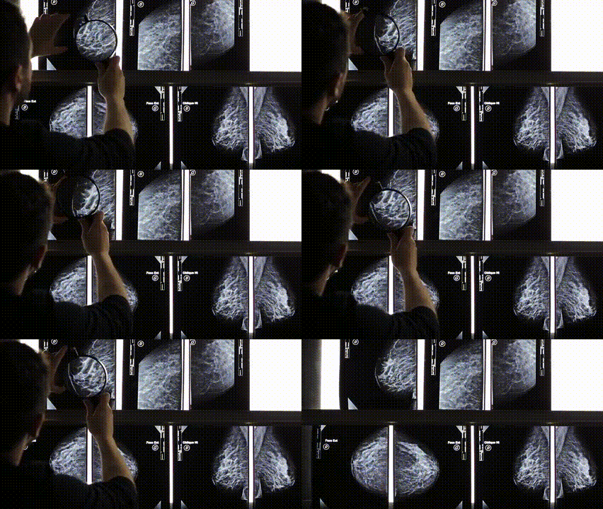
Nowadays, one of the hottest subjects of research in the field of biomedical imaging is digital mammography: this is one of the biggest projects in biomedical image processing and it is conducted by IBM Research. Given the fact that breast cancers are the second leading cause of death for women (accounting for close to 30% of all diagnosed cancers in women), the call for precise and automated detection is crucial. Human image analysis is very difficult, because of the similar aspect of tumors and fat tissues in mammography. Percentages of errors in detection are dramatic, hence the need for an automated system to investigate the suspect areas and the results of which would have high accuracy and high sensitivity, excluding as much as possible instances of false negatives. Such a system would certainly be a major breakthrough in biomedical image analysis. This will be achieved with the use of deep learning, segmentation (instead of only detection) and sophisticated techniques of computer vision and image processing.
Another challenge preventing (or at least slowing) breakthroughs performed by computer vision in medical imaging is the lack of public datasets of the kind of imagenet. That would allow to train systems more thoroughly and to compare over the same dataset models proposed by different teams around the globe. Overcoming this obstacle would facilitate additional innovation and new projects in biomedical imaging. Privacy issues for patients are also a necessary constraint which must be taken into account.

