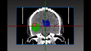
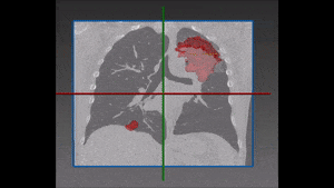
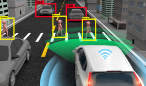
blog
Multiplex IF Analysis
The use of deep learning for analysis of multiplex IF has allowed for a much greater accuracy level for the correct phenotypic classification of cells. When combined with RSIP Vision‘s advanced nuclear detection capability, it allows for the simultaneous analysis of multiple florescent markers on a cell by cell basis. This tool is well suited for multiple applications, especially when using multiple markers to characterize distinct cell populations such as in immune-oncology and IBD.
Circulating Tumor Cells (CTCs)
Circulating tumor cells (CTCs) are rare cancer cells that originate from a tumor and then travel through the patient’s blood or lymphatic system. CTCs have
Detecting Pulmonary Embolism from CT Scan
Pulmonary embolism is a very dangerous condition, which happens when a clot of blood moves from somewhere (generally the legs) to the heart and then
Coronary Arteries Segmentation
Coronary artery disease (CAD) or ischemic heart disease (IHD) has become one of the most common causes of morbidity and mortality worldwide. Patients who suffer
Great Vessels Segmentation with Deep Learning
The great vessels conduct blood to and from the heart. These vessels include the aorta, superior and inferior vena cava, pulmonary arteries and pulmonary veins.
Middle and Inner Ear Segmentation with Deep Learning
Ear pathologies are common in all age groups, and are one of the leading causes for visiting a doctor. In most cases, proper diagnosis can
Larynx Segmentation with Deep Learning
The larynx, also known as the voice box, is a triangular structure in charge of important functions including breathing, voice production and supplying protection to
Liver Tumor Segmentation with Deep Learning
Liver tumors, also known as hepatic tumors, are quite common and some poses a grim prognosis. Therefore, early detection and diagnosis has become a main
Lymph Node Segmentation Module
Lymph nodes are routinely examined and assessed during physical examination of patients in a clinic or hospital setting. Enlarged lymph nodes can be indicators of
Lymph Node Segmentation Module
Lymph nodes are routinely examined and assessed during physical examination of patients in a clinic or hospital setting. Enlarged lymph nodes can be indicators of
Brain Ventricles Segmentation with Deep Learning
Sinus Segmentation with Deep Learning
The paranasal sinuses are air-filled spaces surrounding the nasal cavity. The sinuses include the maxillary, frontal, ethmoidal and sphenoidal sinuses. Due to being air filled, the sinuses make
Brain Hemorrhage Segmentation with Deep Learning
Dental Segmentation with Deep Learning
Dental problems affect people of all ages and ethnic groups, and are common worldwide. With many patients suffering from tooth decay, orthodontic issues and even
Lung vasculature segmentation
Lungs vasculature has a major part in blood oxygenation. The complicated branches of arteries and veins, accompanied by the intricate bronchial tree are in charge
Lung Tumor Segmentation
Lung cancer is the most common cancer related mortality cause among men, and second in women worldwide. Primary lung cancer is usually divided into two
Tissue Analysis with AI
New AI technologies by RSIP Vision are very powerful in analysis of tissues and histopathology. This complex task, which has been haunting for years the medical community, has now a very practical solution: deep learning gives very fruitful results to several challenges, like the segmentation of cells and nucleus and the classification of the cells according to the detected pathologies.
Detection and Tracking of Tumors
RSIP Vision’s oncology software combines detection of lesions and tumors in the human body with tracking those findings along CT scans performed during the research: in particular lung, lymph nodes and liver. These tools enable a quick and accurate assessment of the efficacy of the new treatment.
Automated RECIST Measurement
The golden standard for measuring tumors is the RECIST score. RSIP Vision developed an automated module to accurately measure the RECIST score from CT scans as well as the exact 3D volume of the tumors. Changes in volume are a reliable measure of the progression or remission of the tumor, enabling to evaluate the responsiveness of the treatment in a relatively short time.
Get in touch
Please fill the following form and our experts will be happy to reply to you soon