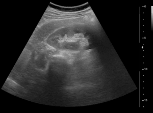
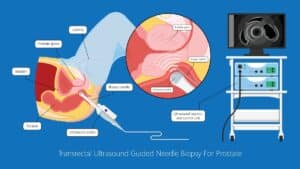
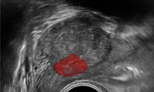
Healthy kidneys have an essential part of proper bodily function. Therefore, early diagnosis and treatment of kidney pathologies is crucial. As people age, some will suffer from a variety of kidney pathologies, most common are kidney stones (nephrolithiasis), but some people may suffer from kidney tumors. Unfortunately, some of these pathologies can greatly affect the quality of life, and some tumors are cancerous. Therefore, many patients need further assessment and treatment. In some cases, contrast enhanced computed tomography (CECT) is indicated for renal imaging. CECT allows the radiologist to evaluate the kidney cortex, medulla, calyces, ureter and vessels. Therefore, kidney segmentation from abdominal computed tomography (CT) images is a prerequisite step towards automated computerized diagnosis of renal pathologies, and treatment of kidney stones including ureteroscopy and percutaneous nephrolithotomy (PCNL). In addition, it is a useful tool for surgical planning of kidney tumors resection and ablation.
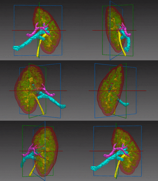
The complexity of kidney segmentation stems from similar density levels of adjacent structures, e.g the liver and spleen and high variability of shape and anatomical position of the kidneys. Number of calyces varies and they have no known tree structure, with a large interpatient variation.
Automated kidney segmentation
RSIP Vision has developed an impressive image processing algorithm for abdominal CT scans that segments the kidney region with high accuracy. This kidney segmentation is also the first step which may enable automatic detection of renal pathologies in the next step.
CT urography in the examination of choice for detecting renal and collecting system pathologies. It consists of four stages – unenhanced, corticomedullary/arterial, nephrographic/parenchymal and urographic phases. Each phase emphasizes different parts of the renal system. The unenhanced phase is used for identifying calcified structures such as stones, corticomedullary allows to detect renal arteries and renal cell carcinoma (RCC), nephrographic phase helps with identifying veins and differentiation between simple cysts and other masses and the urographic phase enhances the collecting system, revealing any malformations or urothelial tumors such as transitional cell carcinoma (TCC).
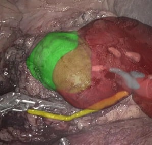
Since kidneys are a relatively small organ, all the tiny details within them must be accurately annotated. In the same line, kidney blood-vessels should be segmented with care. RSIP Vision’s kidney segmentation module is the ideal tool to be used prior to kidney surgical procedures.
A U-Net based neural network was utilized for this task, enabling the learning algorithm to pick up on various important features from within the scan and out-perform any other technique we used, yielding great quality segmentations. Do you have medical segmentation projects? Call us!

 Urology