Denoising
We are posting a series of articles breaking down the automatic detection and quantification of Cystoid Macular Edema(CME). This is a buildup of fluids in the eye near the macula of the eye that can distort vision. We are pioneering a new method for the detection of cysts. We summarized this process in the previous article – Hacking the Automatic Detection Process. Here is the first step of our method in more detail:
Layer Enhancing Denoising
Background
Speckle noise and many small artifacts in the scan might turn layer edge detection into a difficult problem, and due to the small size and shape variability of noise it is hard to devise a non-heuristic method of denoising that will act sufficiently well across different vendors and even across different machines belonging to the same vendor.
On the other hand we can consider using super-pixels as a tool. This is an increasingly popular computer vision concept where neighboring pixels are grouped into coherent units with perceptually shared meaning. Using this concept, super-pixel segmentations are able to find the larger structures of the layers through the noise while being flexible enough to follow each layer’s contours even with heavy disturbances due to pathologies.
Our solution uses this super-pixel segmentation to smooth over the smaller artifacts while introducing larger ones. Afterwards we use a median filter to remove some of the larger artifacts introduced and enhance the layers’ contours.
Essential to the success of the algorithm is understanding the underlying geometric realities of the OCT scans. The usual scan samples the eye with different distances on each axis, i.e. the distance between two adjacent pixels in the scan is different in reality based on their direction with pixels adjacent on the y-axis up to 6 times more distant than neighboring pixels on the x-axis. This means that when smoothing the image using the median filter and a rectangular kernel we are actually being biased towards the y-axis, and thus enhancing each layer’s innate direction.
Input: super-pixel number, kernel size
Algorithm Steps:
- Using the SILC algorithm to segment the image to 35,000 super pixels
- For each super pixel replace its pixels’ intensity with the mean of their intensity
- Filter the image with a 10 x 10 kernel using a median filter
Results:
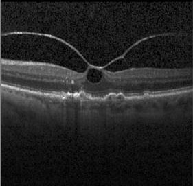
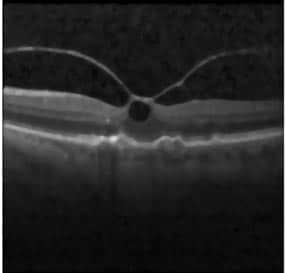
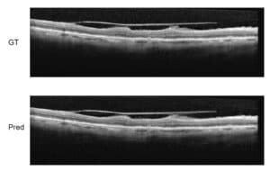
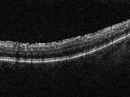
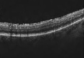
In the above figures it is easy to see how virtually all artifacts inside the retina are discarded, the layers themselves are de-grained and are more consistent which allows easier detection of the change between the layers. N.B. for denoising for layer detection, it not clarity of image that we are trying to achieve clarity regarding where each layer begins and ends.
Notes:
- The SILC algorithm is a popular, efficient and consistent super-pixel segmentation algorithm. More information about the SILC algorithm can be found here: http://infoscience.epfl.ch/record/177415/files/Superpixel_PAMI2011-2.pdf
Curvelet Speckle Denoising
Background
Curvelet denoising is a variant of wavelet denoising where the location in orientation varies with scale and thus is used for multi-scale object representation.
Curvelets are an appropriate basis for representing images (or other functions) which are smooth apart from singularities along smooth curves, where the curves have bounded curvature, i.e. where objects in the image have a minimum length scale, and thus are appropriate for separating speckle noise from actual objects in the images.
Results:
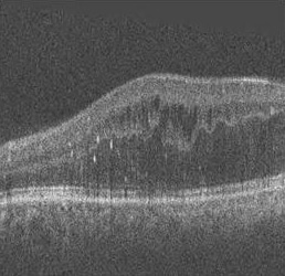
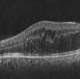
In the above figures it is worth noting the enhancing of the contrast between the cysts in the middle of the scan after the denoising took effect.
Technical notes:
- In this particular implementation this method worked extremely well for Nidek and Topcon machines and made no perceptible changes to images taken with Cirrus and Spectralis machines, which in both cases suggests a connection between the mathematical concept and the underlying noise structure which is worth investigating further in the future.
- Another work that used curvelet transform to denoise OCT retinal scan can be found in the following link and shows the power this method has in speckle denoising: http://www.ncbi.nlm.nih.gov/pmc/articles/PMC2898712/
- More information about the curvelet transform can be found here: J.L. Starck, E.J. Candes, and D.L. Donoho, “The Curvelet Transform for Image Denoising,” IEEE Trans. on Image Proc., Vol 11, No. 6, June 2002.

 Ophthalmology