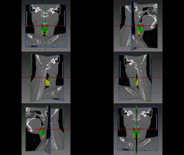
The larynx, also known as the voice box, is a triangular structure in charge of important functions including breathing, voice production and supplying protection to the trachea from aspirations. It consists mostly of cartilages- epiglottic, thyroid, cricoid, arytenoid, corniculate and cuneiform. The cartilages are connected by muscles and connective tissue. The epiglottis that protects the trachea is located at the larynx inlet.

Due to its extraordinary importance, pathologies of the larynx such as laryngeal cancer require thorough care. Treatment options include excision surgery, radiation, chemotherapy or even total laryngectomy. Computed tomography (CT) and magnetic resonance imaging (MRI) are used for staging, surgical planning and follow-up. Unfortunately, the proximity of the larynx to other structures such as nerves and vessels increases the risk for treatment-related complications such as inflammation, nerve damage, fibrosis, tissue breakdown and pain.
Manual review of the scans by radiologists, surgeons and oncologist may be time consuming and prone to errors in staging or treatment choice. Therefore, 3D larynx segmentation for pre-operative planning and for choosing the best sites for radiotherapy, may aid with minimizing these life-altering complications and with creating a favorable outcome.
Automated Larynx Segmentation
RSIP Vision’s scientists engineered a deep convolutional net that segments the larynx, optimizing the tradeoff between accuracy and speed, and at the same time achieving a truly unique solution for this larynx segmentation with deep learning task. Contact us to learn more!

 ENT
ENT
