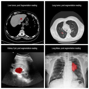
Liver tumors, also known as hepatic tumors, are quite common and some poses a grim prognosis. Therefore, early detection and diagnosis has become a main goal for lowering mortality and morbidity.
Benign tumors include hemangiomas, adenomas, focal nodular hyperplasia (FNH). Although malignant tumors that are found in the liver are metastases of malignancies in other location, primary liver cancer is the sixth most common cancer worldwide, both in developing and industrialized countries.
Prognosis is usually poor, with low survival rates. The most common primary malignant tumor is hepatocellular carcinoma (HCC), and rarely primary tumors are cholangiocarcinoma, sarcoma or hepatoblastoma.
Treatment options include tumor resection or liver transplant. Tumors might also be ablated using radiofrequency or using cryoablation. Moreover, Transcatheter arterial chemoembolization (TACE) or Selective internal radiation therapy (SIRT) are used. In later stages, systemic treatments may be indicated.
Imaging is an integral part of diagnosis, including ultrasound (US), contrast-enhanced computed tomography (CT) and magnetic resonance imaging (MRI). In addition, quality imaging of the tumor aids with choosing the correct treatment protocol and follow-up.
Liver tumors may vary in their location, shape, density, borders. Few tumors may contain calcification, fat or cystic features. Hence, identifying tumors is a difficult task by itself. Moreover, correctly classifying the tumors as malignant or benign can be grueling.
Due to the fact that there is a clear relation between liver malignancy and cirrhosis, these patients need careful and frequent evaluation and imaging in order to minimize mortality. However, differentiating between benign and malignant tumors in patients with cirrhosis can be challenging, due to distorted morphology and liver tissue enhancement. Hence the need for accurate liver tumor segmentation.
Automated Liver Tumor Segmentation
Hence, automated segmentation of liver tumors may help with quicker and more precise diagnosis and follow-up, improve surgical planning and minimize complications during tumor resections and other treatments.
RSIP Vision has developed a deep learning algorithm that reliably detects liver tumors in CT scans, overcoming the challenge posed by the large variance in tumor appearance and location across different patients. The liver tumor segmentation algorithm utilizes a sequential approach, first obtaining a coarse liver segmentation and then using it to perform a tumor segmentation focusing on the liver region. This cutting edge AI model is today’s best solution to liver tumors segmentation. Talk to a Deep Learning expert now!

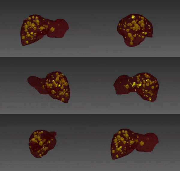
 Medical segmentation