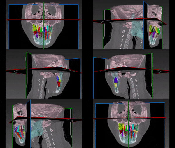
Dental problems affect people of all ages and ethnic groups, and are common worldwide. With many patients suffering from tooth decay, orthodontic issues and even oral cancer, the need of oral surgeries and other treatments increases constantly. Recent advancements in technology allow safer and more accurate procedures, including better preoperative planning aided by 3D segmentation of the teeth and other dental structures.
3D segmentation of intraoral scanners (IOS) computed tomography (CT) dental scans, or cone beam computed tomography (CBCT), which became a useful replacement for regular CT in dental imaging, could be used when planning dental surgeries, such as prosthodontics surgeries including dental implants, root canals treatments, orthodontic treatments, maxillofacial surgeries and even dental biometrics. For instance, automated segmentation could be handy when planning dental implant surgery by allowing the surgeons to better review and inspect the maxilla and its bone quality, which is essential for implant stability. Individual tooth segmentation helps with orthodontic treatment planning and teeth alignment.
Although it is a promising development, automated dental segmentation may turn out to be quite difficult. For example, segmentation of each individual tooth is tricky due to complex teeth arrangements, variation in shape and similarity to adjacent structures such as the tooth socket. The maxilla is also a difficult structure to segment, because of its complex thin bony components including the palate, maxillary sinuses and parts of the orbit walls. The challenge to the segmentation algorithm consists in finding the exact classification in abnormal teeth structures like overlapping teeth and abnormal growth directions.
Automated Dental Segmentation
RSIP Vision’s engineers developed a module for automatic segmentation of the dental structure. It is based on deep learning neural networks and advanced mathematical algorithms from graph theory.
Using those computer vision and artificial intelligence methods, we created a fully automatic and accurate anatomical model of teeth, gums and jaws. Dental professionals use to get an accurate dental and maxillary map in view of specific diagnosis and treatment.

