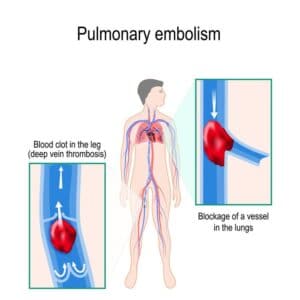
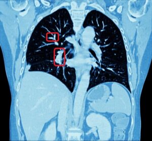
Detecting anomalies in the lower respiratory system requires the use of medical visualizing modalities. Common modalities are e.g 3D sonography, computed tomography, and x ray angiography. The fine scale of the capillary system (reaching as low as 1 nanometer in diameter) imposes difficulties in viewing lung abnormalities, and many times histological samples are taken for in depth analysis. However, edema (leakage) and foreign bulky object, or irregular cell growth can many times be viewed using the mentioned modalities. In such cases, the physicians need to make clinical decision based on prognostic factors integrated from visual and histological information.
Building a lung segmentation software
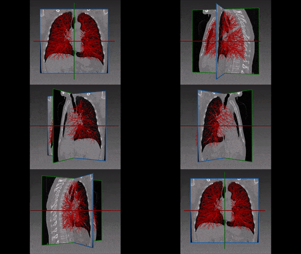
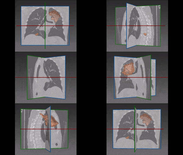
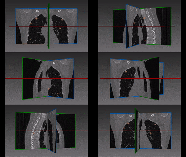
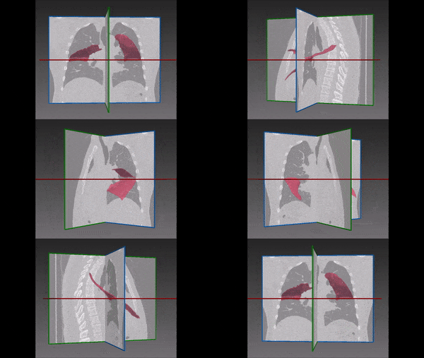
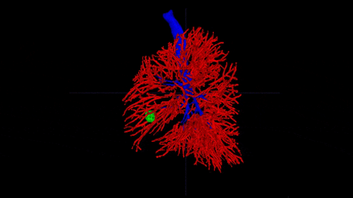
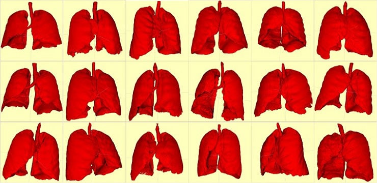
The three dimensional structure of the vascular and capillary tree in the lower respiratory system serves as an exploratory tool for lung segmentation, allowing both diagnosis and planning of invasive intervention. For this end RSIP Vision has constructed an automated segmentation tool to extract blood vessels, the lower respiratory tract, and the pulmonary vasculature. We have utilized advanced segmentation techniques, such as the multi scale Hessian filter and adaptive region growing to segment vasculature from computed tomography images. Our lung segmentation software provides detailed images enabling better clinical analysis and planning. By virtue of our advanced construction, our software can be used to locate bulky tumors (above the detection level), the location of leakage (when applied on X-ray angiography), surgical planing and navigation, diagnosis, and rational treatment plans based on patient specific 3D vascular structure and characteristics.
Read more detailed information about our lung vessel segmentation project. In addition, you can visit our major pulmonary image processing projects page to learn more about RSIP Vision’s activity. Other medical imaging articles are often found in our recommended magazine Computer Vision News.

 Pulmonology