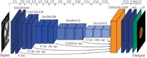
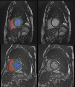
Structural heart diseases include structural deformation of the heart, like valve leakage: the blood flows in two directions and the patient is losing efficiency of the heart. These problems can lead down the line to heart failure. Cardiology offers many possible solutions to structural heart diseases, like implanting a new valve, fix the current valve, fix the valve leaflets and more. When the heart contracts, the leaflets are expected to seal adequately and direct the blood in the desired direction. When there is a malformation or problem with the leaflet, e.g. there may be blood flow to the aorta but at the same time also back into the left atrium – it is something that we want to prevent.
Structural heart procedures are very challenging in that the surgeon is trying to fix the heart structurally without opening the chest cavity. The surgical team tries to accomplish the task using a catheter and images collected via X-ray, Cone-beam CT or any C-arm available, and ultrasound guidance (like transesophageal echocardiogram – TEE).
Artificial Intelligence technology can improve the solutions used by the physician: let’s take as example the mitral valve. The surgeon inserts a catheter, usually in the groin, and advances up in the vena-cava, into the right atrium, through the septum, and starts maneuvering in the left atrium or in the left ventricle. For visualization, the physician uses fluoroscopy and ultrasound. These are usually 2D views, which makes it difficult to know exactly where the catheter is and what its orientation is relative to the existing structure. These limitations are inherent to the imaging modality and even very experienced surgeons can make a mistake. It is very challenging for the doctor to correctly visualize what is going on and they sometimes need to alternate between the ultrasound view and the X-ray view – while the heart is beating and everything around is moving fast. In addition, every decision made at the moment of deploying the device is crucial, because some of the actions have no way back. It is even possible that in a first glance everything looks fine, and only when the catheter is retrieved the medical team learns that something is not ok.
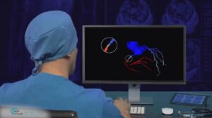
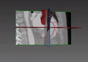
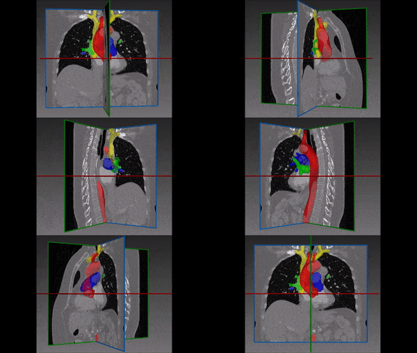
This is where AI can help: all heart procedures have a pre-op imaging done – CT, MRI, or both. Those are 3D views of the heart and they provide an excellent map of the area, which allows the surgical team to plan the procedure accordingly. Of course, these images are not available in real time during the procedure. But advanced deep learning techniques enable to register the X-ray or the ultrasound views on the pre-op CT or MRI (or both). AI can do more: not only register the views, but also correctly label anatomical structures. In this way, the surgeons know that what they are seeing now on the ultrasound is this specific slice of the CT and this 3D position in the CT. At any moment the position of the device is known, as well as labels of the anatomy in the surrounding area. The additional information significantly eases the doctor’s decisions, at a point that one day any doctor (even not a specialist) will be able to perform this difficult task.
At the moment, the companies that develop the medical devices are not the same companies dealing with the imaging modalities. Today, only the surgeon combines this together. It is probably our role – the role of AI companies – to bring up a solution to help the surgeons. The main issue to be solved is data availability. Even though we are talking about a moving organ – the heart – these improvements are already feasible today, thanks to a combination of classic computer vision techniques (segmentation, registration, classification), robotics classic algorithm and deep learning neural networks to get a working solution. Procedures will be faster, more accurate and complications will be significantly reduced. There will not be a single solution for all therapies: each therapy will certainly need a specifically tailored solution.
This is not the final step for AI technology in this area. Robots are increasingly introduced to procedures and delivery systems – and at the same time it is easier to incorporate AI support in robotic procedures. Automatic detection of anatomy and registration of different imaging modalities are close at hand.

 Cardiology