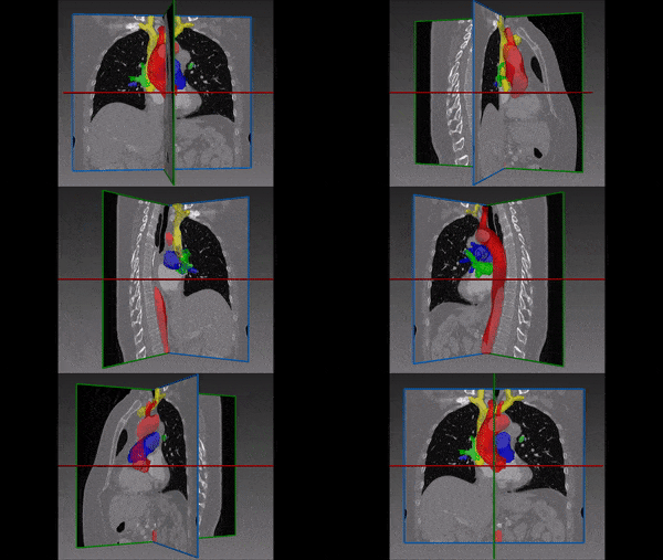
3D Cardiac MRI has an important role in diagnosis, pre-operative planning, and post-operative management of patients with cardiac pathologies. Fully automated segmentation of these images will enable faster and more reliable extraction of data from these images, helping practitioners take more accurate treatment decisions.
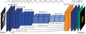
In our project, based on state-of-the-art deep learning techniques, we demonstrate multiclass segmentation of 3D cardiac MRI using a fully convolutional neural network (CNN) with a Unet-based architecture.
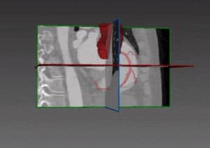
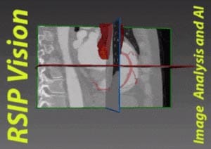
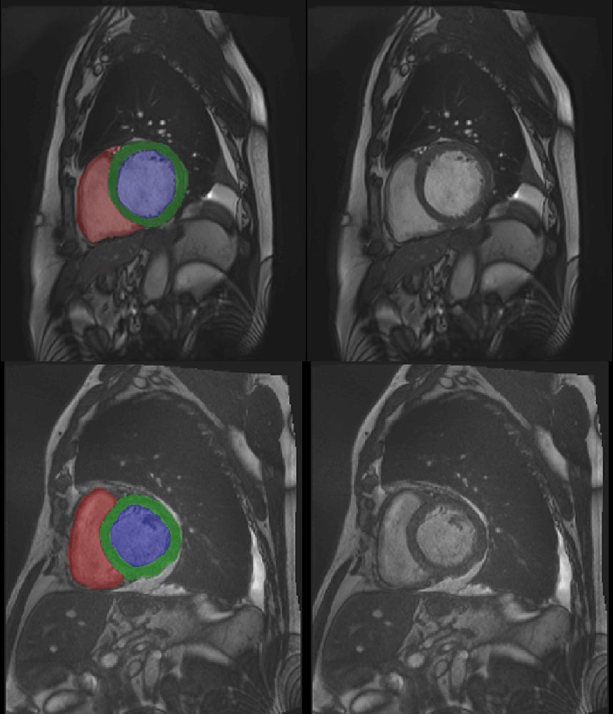
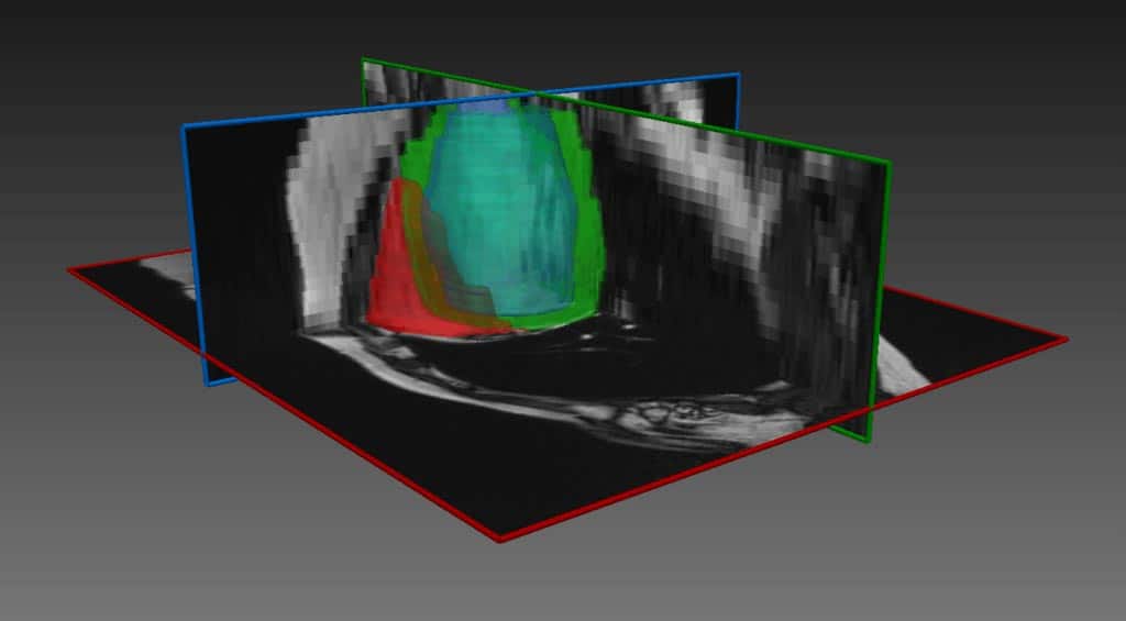
The deep learning network performs automatic segmentation of the right ventricle (red in the image above) left ventricle (blue) and myocardium (green), for both end diastole and end systole phases.
Challenges in 3D Cardiac MRI automatic segmentation
The main challenges of this algorithmic procedure are as follows:
3D data: dealing with 3D data makes for a very large network due to the need for 3D convolution kernels. Since available databases are not very large, this is a major challenge. To overcome it, we have found the correct balance for the size of patches generated from the dataset. They should be small enough to allow sufficient training examples to be generated; but also large enough to contain sufficient information about the region of interest.
Initialization: the size of the CNN also makes it very sensitive to the type of initialization which is used on the convolution kernels. This factor alone can determine if the network will converge or not. We found that the best method in this case was uniform initialization.
Memory – Left and right cardiac ventricles are large, which means that in order to detect them accurately a large input is needed. When GPU memory is limited, it is necessary to find the optimal tradeoff between input size and batch size. One wants to use as large a batch as possible (for normalization reasons), but on the other hand, a large input which can consume vast amounts of memory.
Tackling these challenges, segmentation technology with deep learning by RSIP Vision achieves fast, robust and accurate segmentation of the right ventricle (red) left ventricle (blue) and myocardium (green), for both end diastole and end systole phases. The multiple classes are extracted simultaneously, in constant running time, while the software is easily deployable on standard PC hardware with standard NVidia GPU. Our approach for Artificial Intelligence in cardiology proves to be very effective. The technology presented above for 3D Cardiac MRI automatic segmentation can also be adapted to segment other organs under other imaging modalities, given appropriate training dataset.
Would you like to know how RSIP Vision’s technology and experience can help you obtain better results from your network? Contact our experts now!


 Cardiology